top of page

Fluid Responsiveness

The following algorithm can be used to guide fluid resuscitation using cardiac ultrasound.

Start FR Alg

Do you see the following features on your FoCUS exam?

Hyperdynamic LV

LV ED area <10cm2

SAM

IVC<1cm

Signs of overt hypovolemia (anyone of the following images is considered to be fluid responsive ):

Give Fluid Challenge

Measure VTI or SV before and immediately after 250ml bolus bolus challenge if clinically indicated. Fluid responsive does not mean they need fluid but that they should respond by increasing cardiac output.

FS curve.png

You are probably on the steep portion of the FS curve (above)

Fluid Given?


First Fluid Callenge

Did you observe an increase of the VTI or SV of 15% or more compared to baseline?

Testing

Do not give fluid challenge

You patient is probably on the plateau phase of the FS curve (see below)

Perform passive leg raise to evaluate if patient may benefit from additional volume

AND

Consider other causes of shock: 

-LV/RV failure

-PE 

-Tamponade

-Severe Valve Disease

FS curve.png
Stop fluid challenge

Does your patient have any one of the following:

Spontaneously breathing

Arrhythmia

Spontaneous breaths during intermittent positive pressure ventilation

LV or RV severely dilated or depressed

No signs hypo1

Perform a Passive Leg Raise

Measure VTI or ST before and 1 minute after PLR.

Average over 3 consecutive beats.

PLR.png
Probe Final color_edited.jpg

Did you perform PLR?

PLR

Does your patient have ALL of the following:

Mechanically ventilated

AND

No spontaneous Breaths

AND

TV 8ml/kg

AND

No RV or LV failure

AND

Sinus rhythm

No signs hypo2

Test for Predictors of Fluid Responsiveness

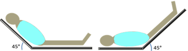
Measure SV or VTI interrogating the LVOT, mitral valve inflow or aortic/carotid flows using PWD or CWD to asses for respiratory variations. Make sure you are scanning as parallel to the flow as possible. 


As an example, on the images above, LVOT PWD with minimal variation. On the right there is observable variation.


Do you observe a variation of  VTI or peak velocity variations >12% 


Test for Respo

References

1. Miller A, Mandeville J. Predicting and measuring fluid responsiveness with echocardiography. Echo Res Pract. 2016;3(2):G1-G12. doi:10.1530/ERP-16-0008

2. Desai N, Garry D. Assessing dynamic fluid-responsiveness using transthoracic echocardiography in intensive care. BJA Education, 18(7):218-226(2018)

bottom of page