
Show me the POCUS
Renal and Urinary Ultrasound
Introduction
Ultrasound is a versatile and inexpensive imaging modality that can be used to characterize acute and chronic renal pathologies. It is considered to be the first-line imaging modality for evaluating azotemic patients to diagnose urinary obstruction. It is effective in detecting renal calculi and vascular pathology. It is also valuable for distinguishing cystic lesions from renal masses. However its complex internal architecture and variable appearance make distinguishing medical causes of renal disease complex. Our goal in this chapter is to use renal POCUS (focused renal ultrasound) to distinguish causes of renal obstruction, calculi and infection.
Relevant Renal Anatomy
The kidneys are retroperitoneal organs with the left kidney situated between the level of the T12-L3 vertebra and usually up to 2cm higher than its right counterpart.
1
3
8
5
2
7
6
4
9
Anatomic structure of the kidney and relevant structures that can be seen on ultrasound. 1. Renal capsule; 2. Nephron within the cortex layer; 3. Renal pyramid; 4. Renal papilla; 5. Minor calix; 6. Major calix; 7. Renal sinus; 8. Renal pelvis; 9. Ureter. Image modified from P Jaworksi CC BY-SA 3.0
Equipment and Technique:
The curvilinear probe (2-5MHz) allows the sonographer to visualize deeper and is the optimal choice of probe to use to visualize the kidneys.
The exam is typically performed in the supine position but due to the kidney's posterior lie in the abdomen, lateral decubitus or even prone may be necessary to optimally visualize the organ. Scanning should occur in a longitudinal plane (or long axis ) and then on its short axis. Deep inspiration causes to diaphragm to displace these structures and move them away from the acoustic shadows cast by the rib cage. Subcostal views can also be used but will not be used as part of the focused renal exam.
Renal artery evaluation has a high failure rate (up to 30%) on expert hands. Intra-renal studies are easier but interpretation of the results are variable which is why Doppler ultrasound is not included as part of the focused renal ultrasound examination.
Kidney Windows
We will be using two windows to interrogate the kidney in B-mode ultrasound. Remember that the evaluation is both in long and short axis. Lets first take a look at the technique before going into the the structures that can be visualized on examination.
Right Kidney
1
Have the patient lie supine with the probe positioned at the mid axillary line. Have the probe marker pointing towards the patient's head. This maneuver should generate a long axis view of the kidney. To improve visualization you might have to ask the patient to take a deep breath to displace the kidneys down to avoid the acoustic shadow generated by the ribs. Rotate the probe 90 degrees from here to get a short axis view of the kidney.


The examination starts at the mid axillary line to evaluate the long axis of the kidney. Probe indicator pointing up as the probe is moved posteriorly.
Left Kidney
2
The left kidney is located more posterior and into the patients back.


The left kidney is examined more posteriorly than that of the right kidney. The lateral decubitus position may be better to visualize the left kidney.
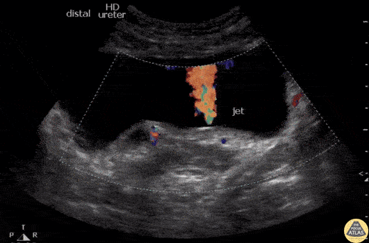
Bladder: ureteral jets
Ureteric jets represent the visualization of normal physiological periodic efflux of urine from the distal end of each ureter into the bladder. As the urine reaches the vesicoureteric junction, it is forced into the bladder after a smooth muscle contraction of the ureter and under ideal conditions, contract twice or more per minute. These jets can be visualized with the use of Color Flow Doppler (CFD) and are seen as a sudden burst of color in the bladder lasting a few seconds .

Ureteral jets. These are seen as shorts burst of color of short duration at the vesicoureteral junction.
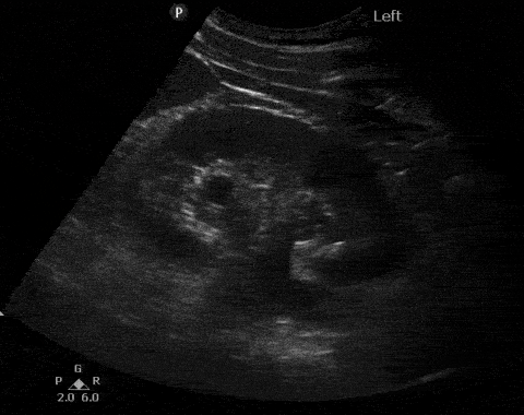
Closer look: sono anatomy of the kidney
The kidney is enveloped in an echogenic capsule which makes this structure easy to identify on ultrasound. The cortex appears isoechoic its echogenicity is very similar to that of the liver or spleen. The medullary pyramids are triangular hypoechoic structures that appear darker than the cortex and the pyramidal shape is not easily visualized on ultrasound. Between the pyramids, the cortex extends in the form of columns (of Bertin). The collecting system is hypoechoic not well visualized unless there is distention. Renal sinus fat is echogenic and occupies a significant portion of the inner kidney.
1
2
3
Anatomic structure of the kidney and its appearance with B mode ultrasound. 1, renal cortex; 2, pyramids, 3, collecting system (minor and major calices and renal pelvis). On ultrasound the cortex has the same echogenic appearance as the liver, the pyramids (renal medulla) is hypoechoic but not anechoic and the renal fat is hyperechoic. You can pause the clip on the right. The collecting system is not well visualized under normal conditions and in fact you cannot distinguish it on this clip. Image modified from P Jaworksi CC BY-SA 3.0
Kidney perfusion

Renal Doppler imaging is via direct visualization of renal arteries/veins. A precise interrogation of the vascular structure of the kidney is beyond the scope of a focused kidney evaluation. However a simple scan can assist in determining if patient with features of hydronephrosis actually has prominent vascular arteries or veins.
Perfusion of the kidney. Depiction of the renal vasculature on the left and on the right a Color Flow Doppler (CFD) window w a low nyquist level to evaluate for both arterial and venous blood. CFD can determine if the ultrasound image seen corresponds to hydronephrosis vs prominent renal vasculature. Image modified from P Jaworksi CC BY-SA 3.0.
Renal Pathologies
The following section displays a range of pathologies that can be seen with performing a focused renal ultrasound examination:
Infection:
Ultrasound (US) is insensitive to acute pyelonephritis as an unremarkable renal ultrasound exam is visible in most cases. The American College or Radiology Appropriateness Criteria for acute pyelonephritis does not recommend imaging for uncomplicated cases. In complicated cases it can offer a low-risk rapid acquisition modality. Some features of abnormal US findings include particulate matter or debris in the collecting system, abnormal echogenicity with edematous (hypoechoic) or hyperechoic (hemorrhagic) areas, gas bubbles or reduced cortical vascularity with the use of Power Doppler.
We can use US to evaluate for complications such as:
Emphysematous pyelonephritis is a fulminant necrotizing infection of the renal parenchyma associated with the formation of gas and predominantly affects patients with diabetes. On US, we see pockets of pair with posterior acoustic shadowing de to the presence of gas.

Emphysematous pyelonephritis. This longitudinal scan of the kidney shows multiple large echogenic foci of air extending into the parenchyma. Notice the acoustic shadow cast by this infection. Image courtesy of SM Almoghazy,MD ( Radiopaedia.org, rID: 88419)
Pyonephrosis is the presence of pus in the kidney and its upper collecting system that can be associated with an obstruction. On US we see the presence of a dilated of the pelvicalyceal system coupled with:
-
Echogenic debris in the collecting system.
-
Fluid filled levels in the collecting system.
-
Incomplete shadows of gas in the collecting system.


Pyonephrosis. Air-fluid levels (arrows) and echogenic debris in the collecting system is seen. Image courtesy of A Dixon MD (Radiopaedia.org, rID: 10432 ) and MS Patel, MD (Radiopaedia.org, rID: 16173)
Renal abscess is the collection of fluid that is infected within the kidney parenchyma. This is typically secondary to complication from acute pyelonephritis. Severe vasospasm and inflammation can result in liquefactive necrosis and its formation.
On US, renal abscess presents as a well-defined hypoechoic area within the parenchyma of the kidney (cortex or coticomedullary region).


Renal abscess. There is a well-defined parenchymal collection with internal echoes (notice the upper marker on the image to the left) and no apparent blood flow through the structure on the image to the right. Images courtesy MS Patel, MD (Radiopaedia.org, rID: 48616)
Renal infarct
Renal infarct appears as an absence of perfusion of the renal cortex using Color Flow Doppler or Power Doppler.

Renal infarct. Notice the absence of perfusion when with the use of Power Doppler. Image courtesy of G Fananapazir, MD (Abdominal Radiology 43(9):1-9)
Hydronephrosis
Hydronephrosis is the abnormal enlargement and distention of the structures of the kidney caused by obstruction of flow. The kidney keeps producing urine but unable to empty creating a buildup of pressure. The most common causes of hydronephrosis include kidney or bladder stones, benign prostatic hyperplasia, cancer (of the kidneys and those related anatomic structures) and urinary retention.
This is the most common indication to use POCUS. Renal POCUS can detect Hydronephrosis with a sensitivity of 77-90% and a specificity of 71- 96% with a positive predictive value 91% compared to CT scans. On imaging we see predictible progressive dilation of structures that continue to balloon because of the buildup of pressure. The structures closest to the obstruction dilate first which implies that the renal pelvis dilates first, followed by the major calyces. Then come the minor calyces. Finally the renal cortex. These compromised structures are filled with pressurized urine and thus appear anechoic on ultrasound and so we focus on the dark areas of the images and its progression to the cortex.
Mild Hydronephrosis (Grade 1):
The first structures to be compromised are those closest to the obstruction, the renal calyces and pelvis (pelviectasis) appear dilated. There is preservation of the cortico-medulla differentiation. Ureters are also not typically seen unless they are distended (hydroureter). Note that normal patients that have not voided for a prolonged amount of time might have these findings.

Comparison side by side of normal appearing kidney on US vs mild hydronephrosis (image courtesy of thepocusatlas.com). Notice the dilated renal pelvis.
Moderate Hydronephrosis (Grade 2):
We now also have dilation of the calyces (caliectasis) on top of the existing pelviectasis. The renal cortex does not present with thinning. As noted by nephropocus.com imagine a black branching tree.


Comparison side by side of normal appearing kidney on US vs moderate hydronephrosis. Notice the hypoechoic collecting system that is in close proximity to the pyramids but does not obliterate them.
Severe Hydronephrosis (Grade 3):
There is gross dilation of the renal pelivis and calyces that compress on the renal cortex. The cortex appears thin with an anechoic "Bear Claw" appearance.


Comparison side by side of normal appearing kidney on US vs severe hydronephrosis. Notice the hypoechoic collecting system that has now obliterated the pyramids and a portion of the cortex
False Positives: Entities that may appear like hydronephrosis on ultrasound include: vascular malformations (which can be confirmed with Power Doppler or Color Flow Doppler), extrarenal pelvis and parapelvic cysts. These are beyond the scope of focused renal US.
Nephrolithiasis
Renal calculi are crystal concentrations formed in the kidney that travel throughout the urinary system. When these excretions are large enough, they can cause obstruction and intense pain. Nephrolithiasis is the most common urinary system condition with up to 12% of the population suffering from it. Calcium is the main composition of these crystals (80%) in the form of calcium oxalate or calcium phosphate. Other types include magnesium ammonium phosphate (struvite), uric acid or cystine crystals.
CT of the abdomen is the imaging study of choice for its diagnosis with a high sensitivity; it also determines size and location. Ultrasonography may be an option as it facilitates radiation-free assessment and is useful for evaluating frequent recurrences, but small stones may be missed.
Determining the size of the stone is imperative as it will dictate if medical vs surgical intervention is warranted. Stones measuring greater than 6mm are less likely to spontaneously pass if they migrate to the ureter. Surgical options are available to those wanting earlier intervention, uncontrolled pain or a solitary functioning kidney.
Ultrasound findings of nephrolithiasis depend on size and location as well as their associated degree of obstruction. Hydronephrosis is a direct consequence of the obstruction by the stone and its absence does not rule out nephrolithiasis since ultrasound is not sensitive enough to rule it out. Here are the other three common findings:
1. Twinkling Artifact.
This is a sonographic artifact created behind the ureteral stones when Color Flow Doppler is used and has a sensitivity of 97% as well as a specificity of 80%. This appears as a multicolored (created by aliasing) high intensity signal and resembles that produced by turbulent flow. This artifact may also present with a comet-tail artifact and leads to a tail like appearance from numerous reverberation signals that the transducer receives and so this artifact makes it seem as though this twinkling artifact goes very deep into the interrogated tissue.
Twinkling artifact. Artifact noted by the blue line. Notice that the ureter having the twinkling artifact also has decreased urine output compared to to the contralateral side. Clip courtesy of the pocus atlas.com
2. Ureteral jets are absent.
Urine does drains at an interval of every 5-10min from the ureters to the bladder at the trigone. This can be confirmed with Color Flow Doppler (CFD) and seen as ureteral jets that empty into the bladder. The CFD scale has to be lowered to 10-20cm/s and what is typically seen is the presence of jets every 5-10min. However in the presence of kidney stones, there is absence of ureteral jets on the obstructed side.

Color Flow Doppler of the ureteral opening at the level of the trigone. We appreciate flow on one of the ureters but not the contralateral side, indicating the presence of a renal stone.
3. Direct visualization of the kidney stone.
We are unlikely to see a kidney stone by direct visualization although it confirms the diagnosis. Most stones lie in the ureter or ureterovesical junction. When seen they appear as hyperechoic structures causing acoustic shadows.


Nephrolithiasis is seen in these two clips. On the left there is a 1x1cm hyperechoic structure on the upper pole of the kidney. On the left, we appreciate the acoustic shadows cast by the crystals.
References
1. Gulati and all. Pictorial Review: Renal ultrasound. Clin Imaging. Sep-Oct 2018;51:133-154. doi: 10.1016/j.clinimag.2018.02.012. Epub 2018 Feb 16.
2. Sibley S, Roth N, Scott C, et al. Point-of-care ultrasound for the detection of hydronephrosis in emergency department patients with suspected renal colic. Ultrasound J 2020; 12(1):31
3. Alelign T, Petros B. Kidney Stone Disease: An Update on Current Concepts. Adv Urol. 2018;2018:3068365. Published 2018 Feb 4. doi:10.1155/2018/3068365
4. Rule AD, Lieske JC, Pais VM. Management of Kidney Stones in 2020. JAMA. 2020;323(19):1961–1962. doi:10.1001/jama.2020.0662
5. Liu N, Zhang Y, Shan K, Yang R, Zhang X. Sonographic twinkling artifact for diagnosis of acute ureteral calculus. World J Urol. 2020 Feb;38(2):489-495. doi: 10.1007/s00345-019-02773-z. Epub 2019 Apr 24. PMID: 31020422; PMCID: PMC6994645.
6. Smith-Bindman, Rebecca, et al. “Ultrasonography versus Computed Tomography for Suspected Nephrolithiasis: NEJM.” New England Journal of Medicine, 18 Sept. 2014