Gastric Ultrasound
Introduction
Perioperative aspiration of gastric contents is a serious complication of anesthesia associated with high morbidity and mortality. Mortality after aspiration pneumonia can be as high as 5% and it accounts for up to 9% of all anesthesia-related deaths. Prevention of perioperative pulmonary aspiration is part of the process of preoperative evaluation and preparation of the patient. The preoperative fasting guidelines were developed to lower the risk of pulmonary aspiration in patients undergoing surgery. They assist health care providers and patients in knowing when to stop eating and drinking. They are considered to be the standard of clinical care and evidence-based practice.
These guidelines however are only intended to a patient population of all ages undergoing elective procedures. The guidelines do not apply to patients who undergo procedures with no anesthesia or only local anesthesia when upper airway protective reflexes are not impaired and when no risk factors for pulmonary aspiration are apparent. The guidelines are also limited in that they may not apply to or may need to be modified for patients with coexisting diseases or conditions that can affect gastric emptying or fluid volume (e.g., pregnancy, obesity, diabetes, hiatal hernia, gastroesophageal reflux disease, ileus or bowel obstruction, emergency care, or enteral tube feeding) and patients in whom airway management might be difficult. These conditions severely limit the applicability to all patients. Finally, the use of cricoid pressure is has been debated since in up to 50% of the population the esophagus is not behind the trachea and thus ineffective when performed. Enter the era of gastric ultrasound.
Gastric ultrasound is a point-of-care tool for aspiration risk assessment. It can determine the nature of the content (empty, clear fluid, thick fluid / solid) and when clear fluid is present, its volume can be estimated. POCUS can be used to measure the gastric antrum at this time since it is the easiest to interrogate. The gastric antrum expands from a baseline empty state as fluid enters the stomach in a linear relation to that of the rest of the body of the stomach up to 300ml. Volumes in excess of 300 ml result in only modest further increases in antral size, with excess volumes being accommodated by more proximal areas of the stomach.
Showmethepocus.com contextualizes Gastric Ultrasound with the following lecture:
Equipment and Technique:
The curvilinear probe (1-8MHz) allows the sonographer to visualize deeper structures at the expense of resolution and is the optimal choice of probe to use to visualize the stomach.
Scanning should occur in a longitudinal plane with the probe held perpendicular to the surface of the skin at the level of the epigastrium as seen in the image below. The patient should be slightly head up at <45 degrees and examined in both the supine and right lateral decubitus position (RLD). The RLD position is going to be the most sensitive at measuring volume. The optimal image should include the left lobe of the liver and the descending aorta. If the descending aorta is not visualized the inferior vena cava should. Set your depth at >4cm and make sure your interrogation sector is not narrow but wide (the piece of 'pie' that the US displays is wide and not narrow).

Diagram of the stomach for reference. Image from Foresight ultrasound.

Optimal probe position for gastric examination. Note that the probe is tilted towards the patient's head.
3D Model of the Gastric Antrum
The clips here are designed to assist you in understanding the structures related to the stomach and also how the ultrasound probe is positioned to provide adequate interrogation of the gastric antrum.
The clips.
Clips 1 through 3 show an animated 3D model of the stomach as it fills with gastric content. Clip 1 is the model with relevant anatomic layers. Clips 2 and 3 represent the same model with an ultrasound sector showing the area of interest during the POCUS interrogation. Clips 3 show what you should be seeing on 2D ultrasound. Clips 2 additionally show the minimal expansion of the gastric antrum as the rest of the stomach fills with additional volume.

1

2

3
A 3D model of the gastric antrum is available here for those interested in using the model in augmented reality.
The clips above were created with the use of Z-anatomy and with the help of F Lakhani, MD
Sono anatomy of the Gastric Antrum
The following images display the normal sonographic features when interrogating the antrum for optimal evaluation. At the very least make sure images are obtained on RLD which increases the volume of the gastric antrum (GA).

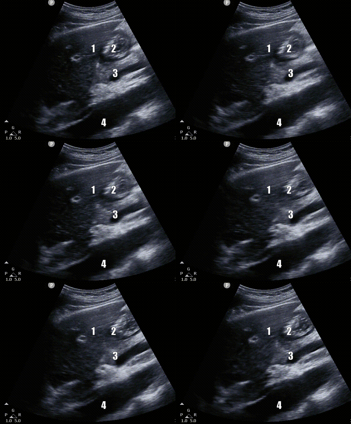
Gastric Sonoanatomy. CT abdomen image showing related structures. On the right, US image with gastric antrum as well as optimal visualization; 1. Liver, 2. Gastric Antrum, 3. Superior Mesenteric Artery, 4. Aorta
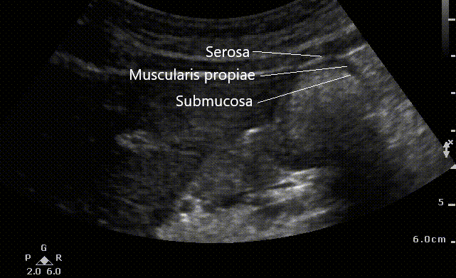
A close up view of the antrum reveals its multiple layers. The gastric wall starts with serosa, muscularis propia, submucosa (seen on the image) followed by submucosa and muscularis mucosae (not seen on this scan). It is important to realize that the measurements to evaluate volume (see below) should include serosa (the outermost hyperechoic layer)
Close up view of the gastric antrum. In this case the stomach appears full and only 3 layers can easily distinguished.
Interrogating Gastric Contents
The following images display a variety of stomach contents from empty to solid food.
Empty GA
Empty GA. Both images have no to very little content within the walls of the stomach. The GA wall layers appear thick
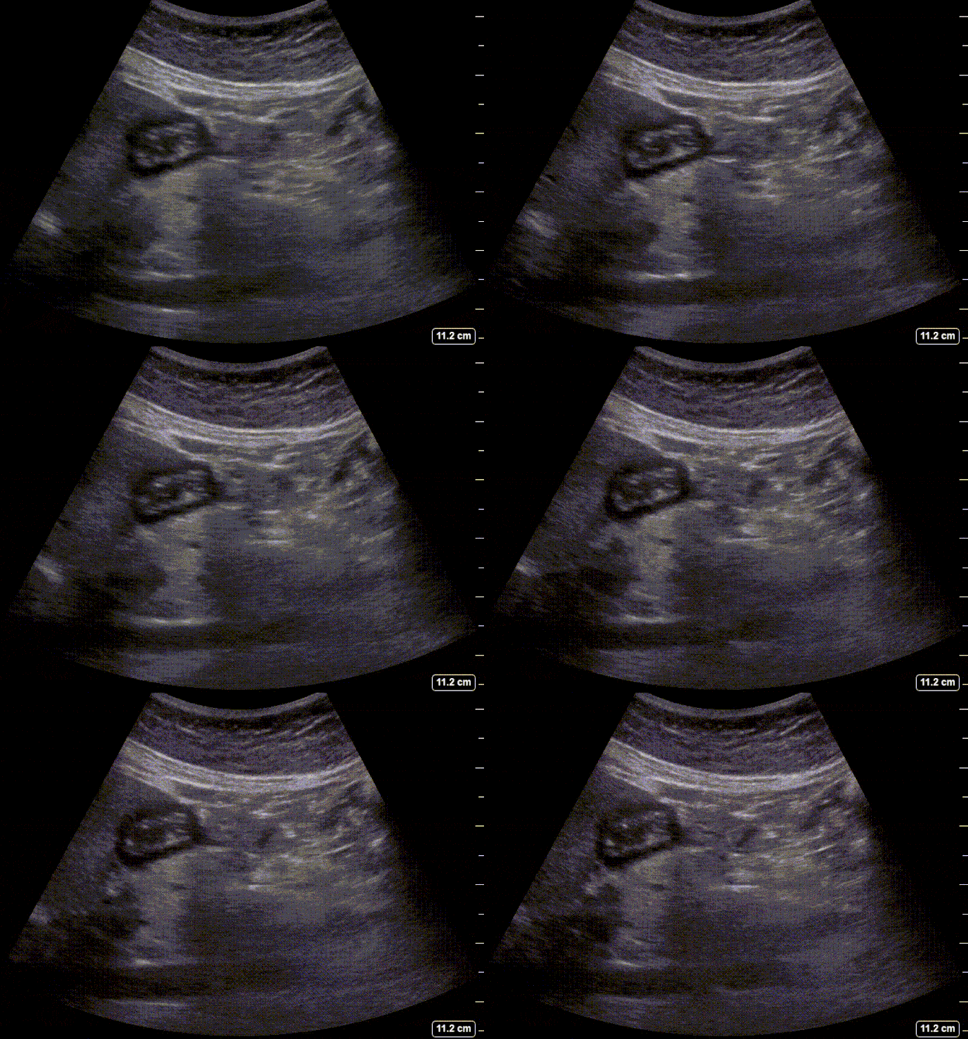
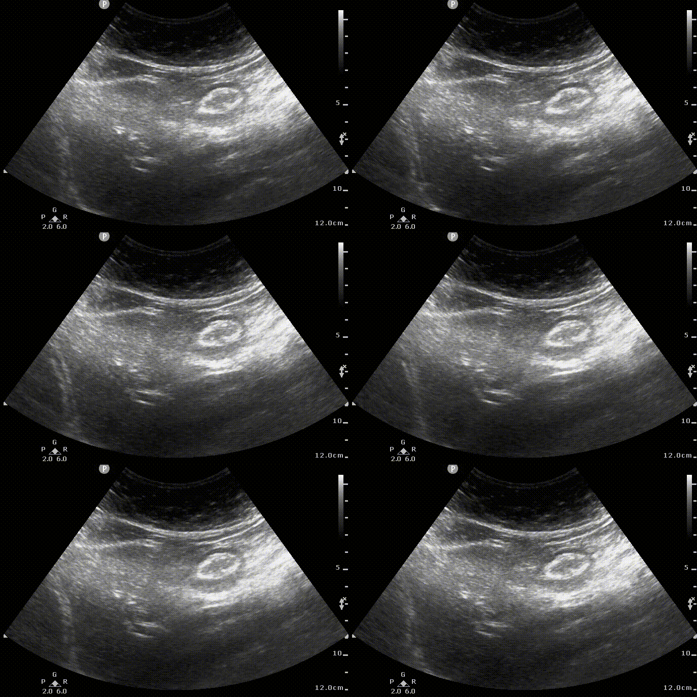
The GA appears flat and has the configuration of a bullseye. When grading the content this would be a Grade 0 GA.
Clear Fluid
Starry night appearance. Occurring shortly after ingestion of clear fluids or effervescent drinks.
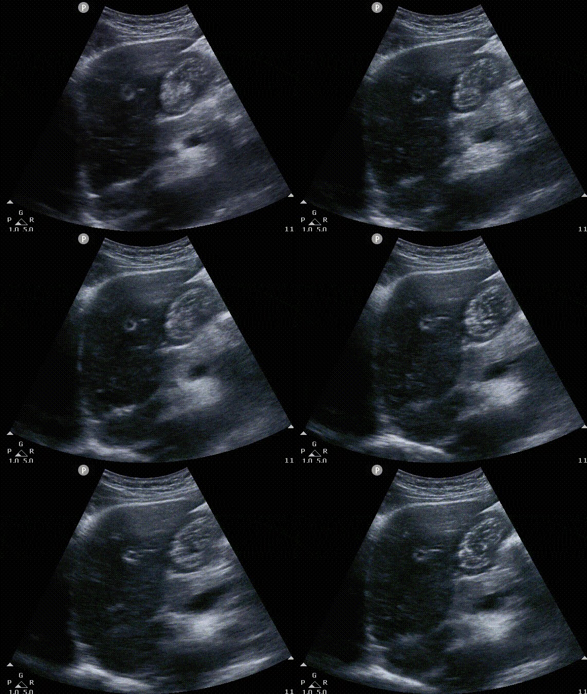
The antrum is clear and distended. Content is anechoic with some hyperechoic debris.
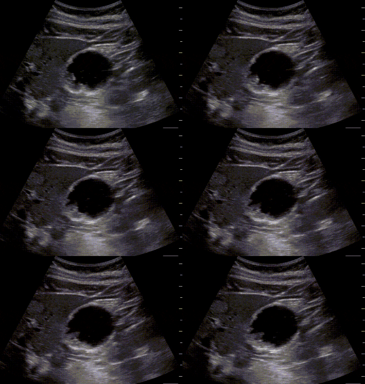
If interrogation yields the above image consider volume assessment to differentiate from baseline secretions and non fasting volume. These images could correspond to a Grade 1 or 2 depending on volume estimation.
Full Stomach

The GA is distended in this image and only its anterior wall is visualized. This is because contents are mixed with air which limits the amount of echo penetration and thus the posterior aspect of the GA is not visualized.
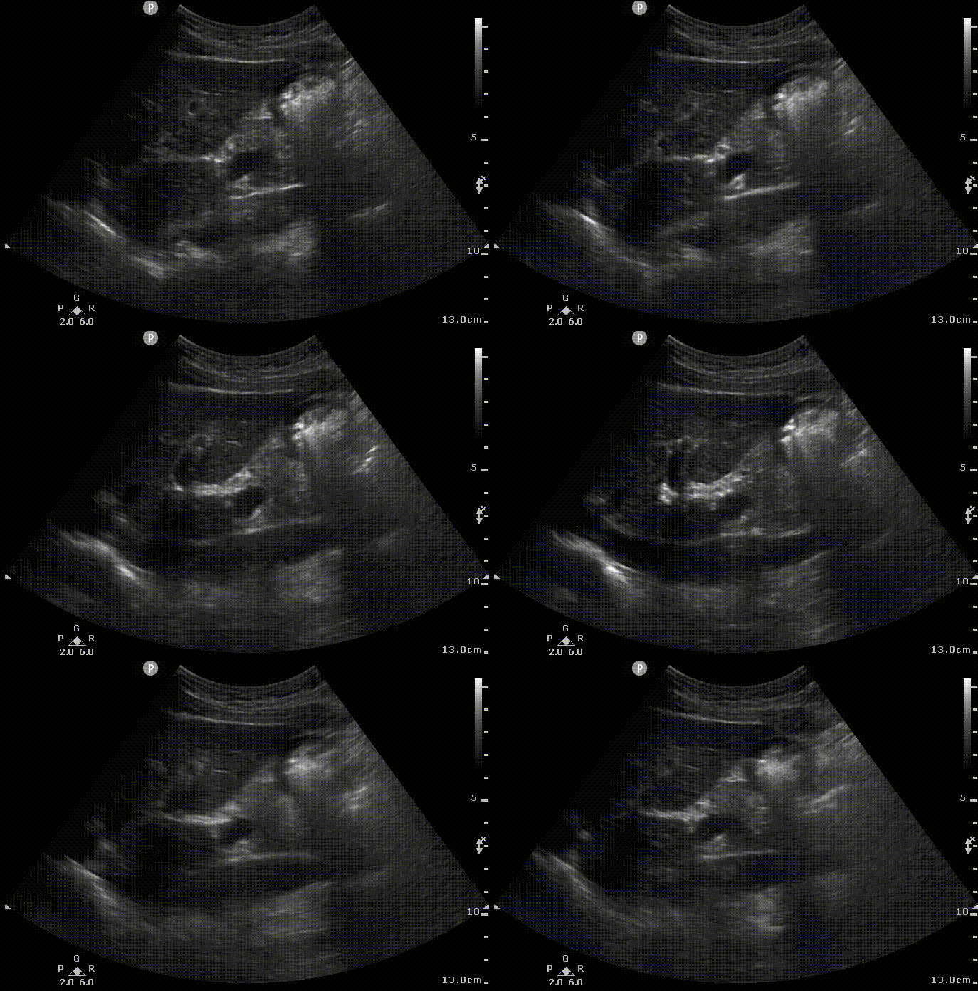
The GA is not as distended but its contents appear with mixed echogenicity.

This is the solid late stage. The borders of the stomach wall are not obscured by air and approx 2h after the meal.
Once you see any of the above images on the supine position you do not necessarily need to have the aorta on view. Or get the patient on the RLD position. These patients are considered as full stomachs.
Volume measurement
The decision making process is simplified for those with Grade 0 (empty stomach) or those with a full stomach. However if there is clear fluid in GA it will be necessary to estimate the volume to make a determination regarding risk.
The volume of the antrum can be measured by tracing the area of this structure from serosa to serosa in the freehand tool on your US machine. It can also be measured by measuring the anteroposterior and craniocaudal diameters.
If the second option is selected the cross sectional area(CSA) = 1/4 x pi x (Anteroposterior x craniocaudal )
Volume (mL) =27 +14.6 x(CSA lateral) - 1.28 X Age

The Cross Sectional area can be either traced or calculated using the craneo caudal distance (cc) and the anteroposterior distance.
Grading system (proposed by Perlas MD and her group )
This antral grading system has been not been validated in children, obese and obstetric patients.
References
1. Practice Guidelines for Preoperative Fasting and the Use of Pharmacologic Agents to Reduce the Risk of Pulmonary Aspiration: Application to Healthy Patients Undergoing Elective Procedures: An Updated Report by the American Society of Anesthesiologists Task Force on Preoperative Fasting and the Use of Pharmacologic Agents to Reduce the Risk of Pulmonary Aspiration. Anesthesiology 2017; 126:376–393 doi:
2. Van de Putte P, Perlas A. Ultrasound assessment of gastric content and volume. Br J Anaesth. 2014 Jul;113(1):12-22. doi: 10.1093/bja/aeu151. Epub 2014 Jun PMID: 24893784.

