Pneumothorax
Pneumothorax is commonly associated with both blunt and penetrating chest injury and is a leading cause of preventable morbidity and mortality. Traumatic pneumothorax, the most common life-threatening outcome in blunt chest trauma, occurs in over 20% of patients with blunt injuries and about 40% with penetrating chest injuries. Its diagnosis is frequently relied on a series of signs and symptoms and x ray findings. Early detection is critical as a delay, specially in those who are receiving mechanical ventilation can lead to progression of the pneumothorax and hemodynamic instability. Enter the era of ultrasound.
Ultrasound has a well stablished role in the diagnosis of a traumatic pneumothorax. The first reported use of ultrasound to detect pneumothorax in humans in 1987. The Focused Assessment with Sonography in Trauma (FAST) examination has now been modified to include lung imaging as part of the evaluation in a trauma patient and now called E-FAST examination, with ‘E’ standing for extended views.

Pneumothorax. Image from Foresight ultrasound.
Based on international recommendations, lung ultrasound should be used in clinical settings when pneumothorax is in the differential diagnosis. It more accurately rules in the diagnosis of pneumothorax than supine anterior chest radiography. Lung ultrasound more accurately rules out the diagnosis of pneumothorax than supine anterior chest radiography.
In extreme emergency, absence of any movement of the pleural line with lung sliding or lung pulse, coupled with absence of B-lines allows prompt and safe diagnosis of pneumothorax without the need for searching the lung point as we will discuss here.
Here we present findings suggestive of pneumothorax. To view the integration of these findings in context we suggest going through the pneumothorax algorithm here:
Equipment and Technique:
A linear high frequency probe (5-13MHz) may be adequate enough to analyze superficial structures such as the pleural interface of the lung and give optimal resolution. Other probes used to interrogate the pleural space include the curvilinear probe (1-8MHz) that allows the sonographer to visualize deeper structures at the expense of resolution. Even phase array transducers used in cardiac imaging (2-8MHz) can be used to view pleural space.
The air in the chest cavity will tend to rise to the least dependent area. In a supine patient interrogating an anterior chest at the second to fourth intercostal spaces. The interrogation should proceed in a systemic fashion as seen no pneumothorax in a region does not obviate its presence in other regions.
Sonographic signs of pneumothorax:
1. Absence of lung sliding. A-lines
When air is present between parietal and visceral pleura, visualization of the normal pleural sliding is absent. M mode can also be used to confirm lack of lung sliding. This tracing in a pneumothorax will only display one pattern of parallel horizontal lines above and below the pleural line and thus have a stratosphere or barcode sign. The absence of lung sliding does not necessarily indicate that a pneumothorax is present.
Lung sliding is abolished in the following conditions:
-Acute Respiratory Distress Syndrome (ARDS)
-Pleural adhesions
-One lung ventilation
-Pulmonary fibrosis
-Atelectasis
-Phrenic nerve paralysis
Presense of A-lines on US
These reverberation artifacts appearing as equally spaced repetitive horizontal hyperechoic lines reflecting off of the pleura.


A-lines appearance on 2D ultrasound. On the left a still image. In the middle clip we appreciate a still pleural line and absence of lung sliding. On the right M mode showing barcode sign on the same interrogated segments of the lung.
2. Absent B-lines
The presence of air in the interface between parietal and visceral pleura obliterates B lines as it hinders the propagation of sound waves. The negative predictive value is 98–100% which implies that visualization of even one comet-tail essentially rules out the diagnosis of a pneumothorax. When present, B- lines obliterate A lines.
B- lines present on this clip. Its presence rules out a pneumothorax.

3. Absent Lung Pulse
Lung pulse is the rhythmic movement of the pleura in synchrony with the cardiac rhythm. Its presence indicates that the parietal and visceral pleura oppose one another, and so its presence rules out a pneumothorax. To visualize a lung pulse have your probe interrogate the pleura close to the sternum where the internal thoracic artery lie.
Lung Pulse. Its presence rules out a pneumothorax.

Comparison: lung sliding vs lung pulse
Before we get into the US sign that confirms a pneumothorax, lets review these two here:


Lung sliding vs pulse. On the left, we appreciate the movement of the pleural line with respiration. On the right, the pleural line seems pulsating at a rate of 70 bpm. Both of these ultrasound findings exclude a pnuemothorax.
4. Lung Point
This sign is pathognomonic of a pneumothorax and it is a finding occurs at the border of a pneumothorax. It is due to sliding lung intermittently coming into contact with the air that has accumulated in the chest. Essentially you see the air because lung sliding stops from occurring. With M-mode you will visualize alternating ‘seashore’ and ‘stratosphere’ patterns are depicted over time. However even with a large pneumothorax you may not see a lung point. See the illustration at the introduction of this chapter for an anatomical reference guide.
In extreme emergency, absence of any movement of the pleural line with lung sliding or lung pulse, coupled with absence of B-lines allows prompt and safe diagnosis of pneumothorax without the need for searching the lung point as we will discuss here.

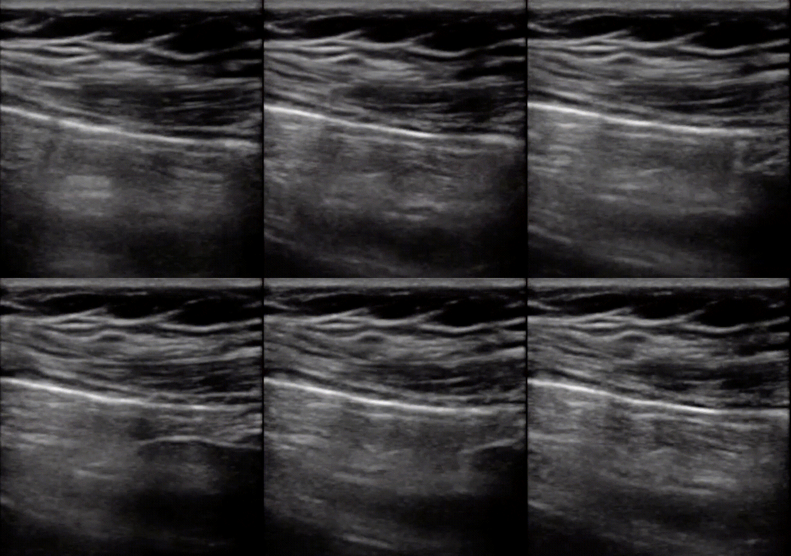
Pneumothorax. Lung point can be seen here as the border between normal pleura and air. Image courtesy of Foresight ultrasound.

1

2
Lung Point seen on 2D and M mode using a linear probe. 1 , the normal pleural line is seen originating form the right and moving towards the left. In it's absence we see A-lines and thus it is this interface between lung sliding and air. 2, M-Mode image showing the interface of air (left) and normal pleura (right) separated at the red dot on the screen.

Lung Point is seen on this 2D scan using a phased array (cardiac) probe.
You can use the following algorithm to guide you in your differential diagnosis to rule out or confirm a pneumothorax:
References:
1. Husain L and all. Sonographic diagnosis of pneumothorax. J Emerg Trauma Shock. 2012 Jan-Mar;5(1):76-81
2. Volpicelli G and all. International evidence-based recommendations for point-of-care ultrasound. Intensive Care Med. 2012 Apr;38(4):577-91.doi: 10.1007/s00134-012-2513-4. Epub 2012 Mar 6.
