
Show me the POCUS
Surface Cardiac Ultrasound- The Planes
In this small section we will be going over the spatial representation of the cuts made by the cardiac probe on the FoCUSed exam.
Introduction
The ultrasound machine extrapolates a 2D image on a 3D object. If structures are not lined up correctly your ultrasound assessment may be incomplete or you may not make out any actionable information from the clips you have obtained. This is is a very visual section in which we will be using a 3D heart model and make the cuts needed for a FoCUSed exam.
Animated Heart Model


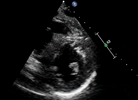
1. Parasternal Short Axis


RV
LV Ant
Inf
Lat
3D heart model seen from different angles with scan sector interrogating the short axis of the LV. The plane cast by the ultrasound beam appears from left to right on the corresponding clip. Corresponding mid papillary view seen on the clip on the right lower corner. RV, right ventricle; LV, left ventricle; Ant, anterior wall; Lat, lateral wall; Inf, inferior wall.
By moving the plane more proximal from the mid papillary short axis we get the parasternal short axis at the mitral valve level and at the aortic valve level.

RV
PV
TV
IAS
AV
Moving the probe towards the head or feet changes the view you see on the parasternal short axis view. TV, Tricuspid valve; RV, Right Ventricle; PV, Pulmonic valve; AV, Aortic valve; IAS, Interatrial septum; MV, Mitral valve.
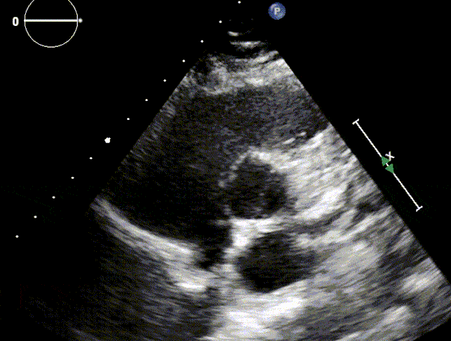
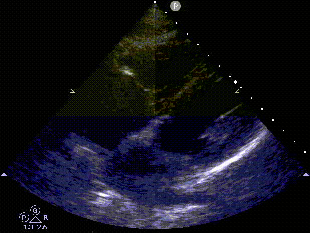
2. Parasternal Long Axis


MV
Left Atrium
LV
AV
LVOT
RV
3D heart model seen from different angles with scan sector interrogating the long axis of the heart. The plane cast by the ultrasound beam appears from left to right on the corresponding clip. On the right lower corner, the representing 2d clip. LV, Left ventricle; RV, Right ventricle; AV, Aortic valve; LVOT, Left ventricle outflow tract; MV, Mitral Valve.
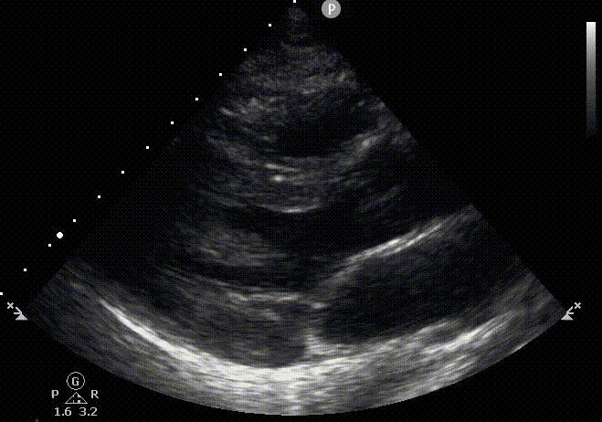
3. Apical 4 chamber


RV
TV
LV
MV
Left Atrium
IAS
3D heart model seen from different angles with scan sector interrogating the apical 4 chamber view of the heart. The plane cast by the ultrasound beam appears from left to right on the corresponding clip. 1. Parasternal short axis; 2, Parasternal long axis. 4 Subcostal View; 5 IVC view. LV, Left ventricle; RV, Right ventricle; AV, Aortic valve; IAS, Inter atrial septum; MV, Mitral valve; TV, Tricuspid valve.
3. Apical 4 chamber To 5 chamber view


4. Subcostal view


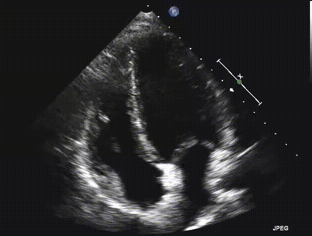
5. Inferior Vena Cava view


IVC
RV
Right atrium
TV
3D heart model seen from different angles with scan sector interrogating the IVC view. The plane cast by the ultrasound beam appears from right to left on the corresponding clip. IVC view. Marker directed towards the sternal notch. RV, Right ventricle; IVC, Inferior vena cava; TV, Tricuspid valve.
References:
1. Zimmerman JM, Coker BJ. The Nuts and Bolts of Performing Focused Cardiovascular Ultrasound (FoCUS). Anesthesia and analgesia 2017; 124: 753-760.
2. Spencer KT, Kimura BJ, Korcarz CE, Pellikka PA, Rahko PS, Siegel RJ. Focused cardiac ultrasound: recommendations from the American Society of Echocardiography. J Am Soc Echocardiogr. 2013 Jun;26(6):567-81. doi: 10.1016/j.echo.2013.04.001. PMID: 23711341.