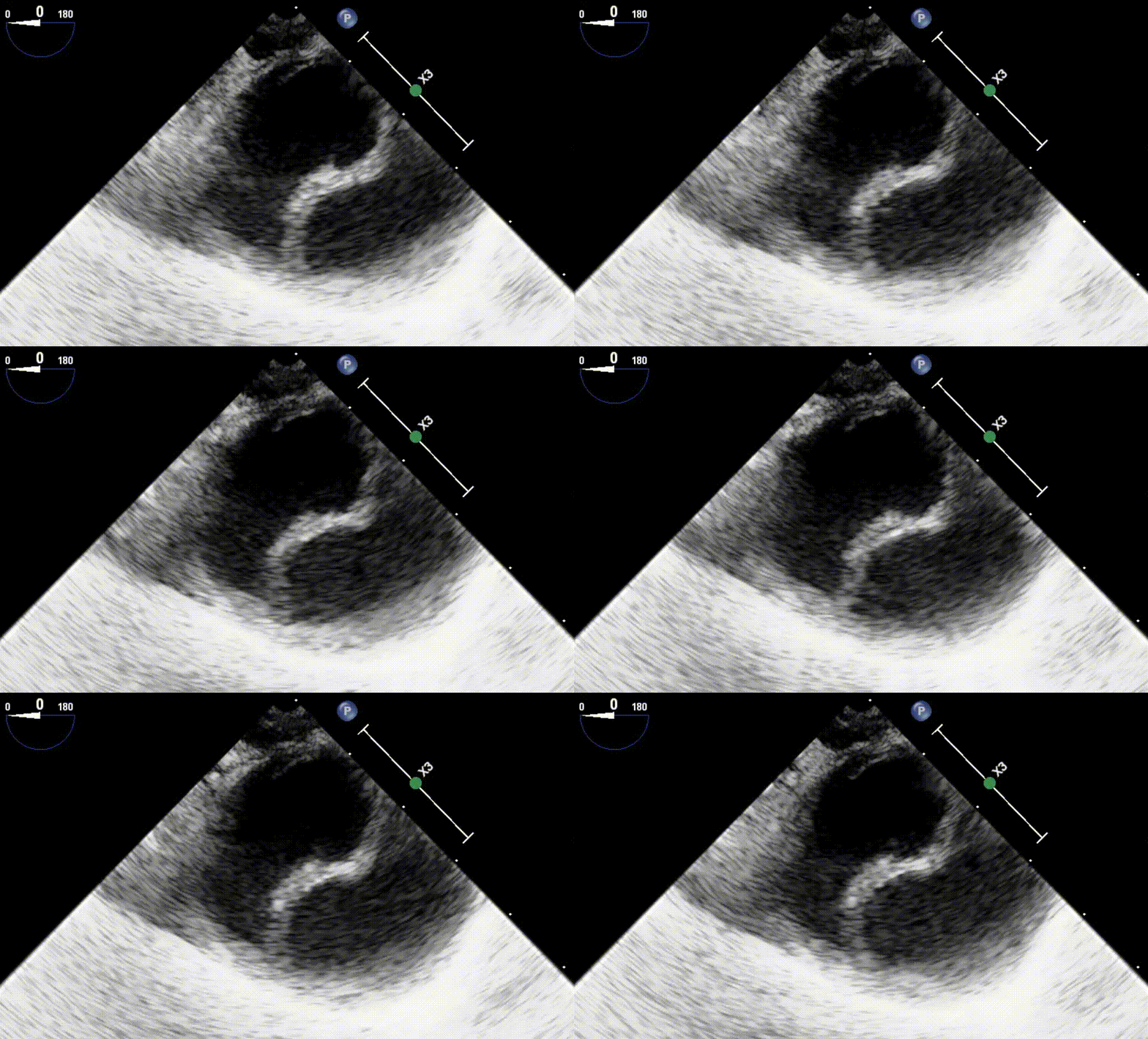
Mid Esophageal 4 Chamber View
To obtain this view the TEE probe is advanced to the
mid-esophagus behind the LA.
The sector depth should be 14cm and omniplane of 0-10°.
Clockwise:
Clip 1. Normal LV and RV function.
Clip 2. Severely Depressed RV and hyperdynamic LV.
Clip 3. Severely depressed LV and RV function.
Clip 4. Flail of the posterior MV leaflet indicating Severe Mitral Regurgitation

RA LA
TV AMVL PMVL
IS AL
RV LV
1
2
3
4
Here we visualize: Left Atrium (LA), Right Atrium (RA), Left Ventricle (LV) [inferoseptal (IS) + anterolateral (AL) walls], Right Ventricle (RV), Mitral Valve [anterior(AMVL), posterior (PMVL) leaflets], Tricuspid Valve [Septal (STVL), Anterior (ATVL) leaflets]
Mid Esophageal 2 Chamber View
To obtain this view, from ME 4 chamber omniplane to 90°
On these images,
Clip 1. Normal Function.
Clip 2. Severely Depressed LV EF. RA also appears enlarged on this view.

LA
PMVL AMVL
Inferior Anterior
LV
Here we visualize: Left Atrium (LA), Left Ventricle (LV) [Anterior and Inferior walls], Mitral Valve [anterior(AMVL), posterior (PMVL) leaflets], coronary sinus (CS).
Mid Esophageal Long Axis
To obtain this view, from ME 4 chamber omniplane to
120-130°
On these images,
Normal function vs
Stuck non coronary or left coronary cusp.

LA
PMVL AMVL
LVOT AV
Inferolat
Antero sept
LV
Here we visualize: Left Atrium (LA), Left Ventricle (LV)[anteroseptal and inferolateral walls], Mitral Valve [anterior (AMVL), posterior (PMVL) leaflets], Aortic Valve (AV), Left Ventricular outflow tract (LVOT).
Mid Esophageal Ascending Aorta Long Axis
To obtain this view from ME 4 long axis withdraw the probe to bring the right pulmonary artery in view.
On these clips and clockwise:
Clip 1. Normal Findings.
Clip 2. A drop off is visualized mid ascending aorta highly concerning for dissection.
Clip 3. A dissection flap is visualized.

Here we visualize: Ascending Aorta in Long axis, Right pulmonary artery in short axis.
Mid Esophageal Ascending Aorta Short Axis
To obtain this view from the ME Ascending Aorta Long axis omniplane to 0 degrees.
On the following clips and going clockwise:
Clip 1. Normal findings.
Clip 2. Dilated ascending aorta on short axis with dissection flap.
Clip 3. Saddle Pulmonary embolism.
Clip 4. Dissection flap on ascending aorta. B, true lumen while A and C are false lumens.

RPA
SVC
Ao
MPA
1
2
3
4
Here we visualize: Main Pulmonary Artery (PA), Right Pulmonary Artery (RPA), Ascending Aorta in short axis (Ao) and Superior Vena Cava (SVC).
Trans Gastric Mid Pappillary Short Axis
To obtain this view, from the ME 4c view advance the probe to the stomach then anteflex to contact stomach wall and inferior wall of heart.
Going row wise:
Clip 1. Normal EF function.
Clip 2. Akinetic walls: anterior, anterolateral, inferolateral.
Clip 3. Hypokinetic walls: anterior and anteroseptal .
Clip 4. Global LV Depression w severe left ventricle hypertrophy.
Clip 5. Severe Global LV Depression.
Clip 6. Circumferential pericardial effusion and hyperdynamic LV

Inf
Inf sept Inf lat
Ant sept Ant Lat
Ant
1
2
3
4
5
6
Here we visualize: LV [ Mid anterior, mid inferior, mid anteroseptal, mid inferoseptal, mid anterolateral, mid inferolateral walls], anterolateral and posteromedial papillary muscles, Right Ventricle.
Mid Esophageal RV inflow outflow
To obtain this view from the ME 4c omniplane to 60 to 70 degrees.
On the first clip we see a stuck left coronary cusp. The other clip was taken from a patient with severe biventricular depression.

LA
IAS
RA
TV PV
Here we visualize: Aortic Valve (AV), Tricuspid Valve (anterior/septal and posterior leaflet ), Left Atrium (LA), Right Atrium (RA), Pulmonic Valve (PV), Right ventricular outflow tract (RVOT).
Mid Esophageal Aortic Valve Short Axis
To obtain this view, from the ME RV inflow outflow view, center on the Aortic Valve.
We see aortic stenosis on both clips. The first clip with all cusps severely sclerosed and limited excursion. The other with the non coronary cusp stuck.

LCC
NCC
RCC
Here we visualize: Aortic Valve (AV)[Non coronary cusp NCC, Left coronary cusp LCC and Right coronary cusp, RCC], Tricuspid Valve (anterior/septal and posterior leaflet ), Left Atrium (LA), Right Atrium (RA), Pulmonic Valve (PV), Right ventricular outflow tract (RVOT).
Mid Esophageal Bicaval
To obtain this view from the ME 2 chamber view rotate the probe right until the IVC and SVC can be seen.
The first clip has normal findings although we cannot fully interrogate the IVC . On the other we see a secundum ASD with the use of Color Flow Doppler on this modified bicaval view (since we see the tricuspid valve)

LA
IAS
IVC
SVC
CT
Here we visualize: Left Atrium (LA), Right Atrium (RA), Inferior Vena Cava (IVC), Superior Vena Cava (SVC), Inter-atrial septum (IAS), Crista Terminalis (CT).
Mid Esophageal Descending Aorta Long and Short Axis
To obtain this view from the ME 2 chamber view rotate the left to find the aorta. The long axis view will be seen orthogonal to this.
The first clip has both views taken at orthogonal (90 degree) planes from each other. They display high degree of calcification within the lumen. The other clip shows a dissection flap.


Here we visualize: Descending Aorta
References
1. Hahn R and all. Guidelines for Performing a Comprehensive Transesophageal Echocardiographic Examination: Recommendations from the American Society of Echocardiography and the Society of Cardiovascular Anesthesiologists. J Am Soc Echocardiogr 2013;26:921-64


