Regional Perfusion of the LV
To simplify this challenging topic we go over understanding what segments are and corresponding coronary perfusion territories. We then need to understand the geometric views and their corresponding segments since we need to be able to locate these regional wall abnormalities in at least 2 cardiac windows. Then we can finally analyze a segment and determine the degree in which it appears compromised.
To assess regional wall motion of the LV, we divide it into segments. These segments reflect coronary perfusion territories. This result in segments with comparable myocardial mass, and allow standardized communication within echocardiography and with other imaging modalities. Regional myocardial function is assessed on the basis of the observed wall thickening and endocardial motion of the myocardial segment.
There are various segmentation models availabe and we will be looking at the 17 segments used in regional wall assessment and out of the scope for FoCUS interrogation since multiple chamber views are needed. However they are mentioned here for illustration purposes
As a reminder, the objectives of FoCUS here is to do semiquantification of ranges of function (e.g., the left ventricle is hyperkinetic, normokinetic, hypokinetic, or severely hypokinetic).
1A. Coronary perfusion model: 3D overview
The right coronary artery (RCA) originates from the right aspect of the aortic bulb. The RCA gives of the Right Marginal Artery (RMA) which supplies the entire right ventricle. The RCA gives off the Posterior Descending Artery (PDA).
The left main coronary artery branches into the left anterior descending artery (LAD) and the left circumflex coronary artery (Cx). The LAD gives off the Diagonal (D1) branches which supply the anterior and anterolateral walls of the left ventricle. The Cx give off the obtuse marginal arteries (M1) which supply the lateral wall of the left ventricle
3D model showing major coronary arteries. Images created with the use of Z-anatomy.
1B. Coronary perfusion territories
Although certain variability exists in the coronary artery blood supply to myocardial segments, segments are usually attributed to the three major coronary arteries. The LV is divided into base mid and apex while on the parasternal short axis view. The following images represent our current model of perfusion.

Base


AL
PM
Mid
RCA
Circ
LAD

Apex
Coronary perfusion territories. On the right, the cuts obtained depending on the level of the short axis; base, mid or apex. We will concern ourselves with the midventricular walls for a focused cardiac assessment. Notice that the anterolateral (AL) papillary muscle is perfused by two vessels while the posteromedial (PM) is only perfused by one in this model. RCA is the right coronary artery, the LAD is the Left anterior descending artery and the Circ or Cx is the Circumflex artery.
2. Segmentation model and Orientation
The LV is divided into base, mid and apex while on the parasternal short axis view. Regional wall can thus occur at each of the segments walls.


The full 17 segmentation model is seen here. For the purposes of FoCUS we will be concerned on the base and mid segment of the 2D cut here depicted in gray and red. On the right, a mid pap short axis view and their corresponding segments. You will not have to memorize the numeration of the segments but their relative location to each other so you can correlate patterns of prefusion and segments.
Lets look at the perfusion of the segments that can be analyzed with the Apical 4 chamber view and Parasternal long axis for completness:

Apex
Mid
Base
Ant Lat
Cap
Inf sep
Base
Mid
Apex
Inf lat
Ant sep
Parasternal long axis and apical 4 chamber view with labeled segments. 1, Parasternal long axis and 2, Apical 4 chamber view. Inf sep; infero septal, Ant lat; anterolateral; Ant sep: antero septal and Inf lat, inferolateral wall.
Remembering the segments on the parasternal short axis model appears easier to do when compared with the other two views so lets make a side by side comparison of the short axis with those others so we can have spatial awareness of their perfusion segments. Lets start with the Apical 4 chamber view.

Mid
Mid papillary walls and corresponding apical 4 chamber segments. The colors on these clips correspond to the same walls. Red, mid papillary inferoseptal wall and in white, mid papillary anterolateral wall. Notice that the bisecting purple line is actually the cut that the ultrasound probe displays on the apical 4 chamber view. Based on the current model the Red colored wall is perfused by the RCA and LAD while the white wall, LAD and Cx.
Now a side by side of the short axis view with the long axis view:

Mid papillary walls and corresponding parasternal long axis segments. The colors on these clips correspond to the same walls. Purple, mid papillary anteroseptal wall and in white, mid papillary inferolateral wall. Notice that the bisecting red line is actually the cut that the ultrasound probe displays on the parasternal long axis view. Based on the current perfusion model the Purple colored wall is perfused by the LAD only while the white wall, the RCA and Cx.
3. Regional Wall Scoring System
For the purpose of FoCUS we will only be using 4 views (excluding the IVC view) which severely limits our ability to look at all the segments of the wall and we focus on visualizing major abnormalities i.e. severely hypokinetic or dyskinesis.
The recommendation is that each segment be analyzed individually and in multiple views. A semiquantitative wall motion score can be assigned to each segment to calculate the LV wall motion score index as the average of the scores of all segments visualized. Regional myocardial function is assessed on the basis of the observed wall thickening and endocardial motion of the myocardial segment. Regional myocardial function is assessed on the basis of a. observed wall thickening (in systole as compared to diastole) and b. endocardial motion of the myocardial segment with wall thickening being the most important element.
Table 1 showing the regional wall scoring system when evaluating regional wall motion abnormalities based on latest ASE recommendations. Percent increase in thickness refers to the change of wall thickness observed in systole as compared to diastole.
Memorizing the grade of regional wall dysfunction is not part of FoCUS. Understanding the percentage change in all thickness is an important consideration when assessing a region.
4. Major perfusion defects
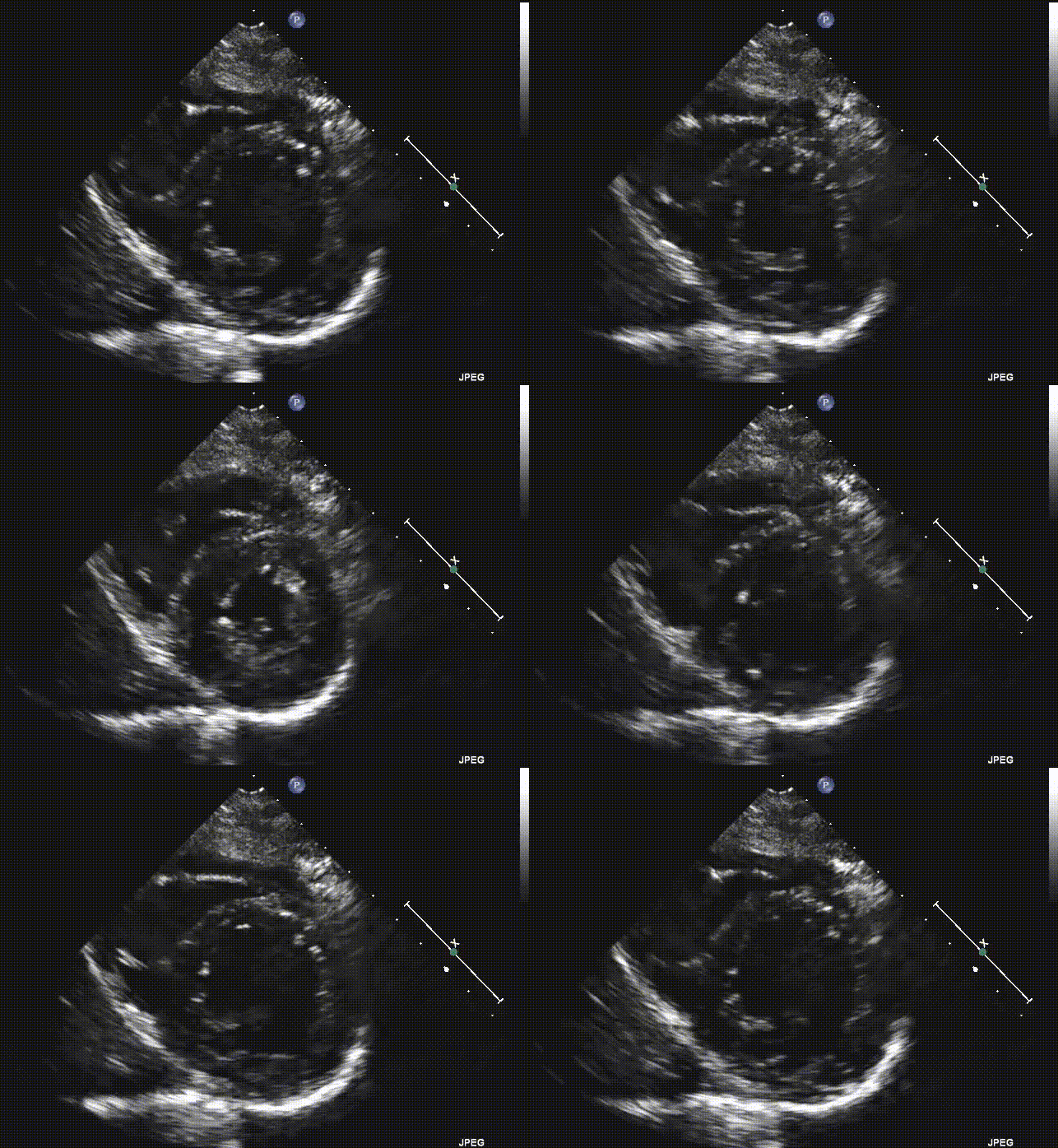
In an effort to recognize major wall abnormalities the following are comparisons to a normal wall motion.
Now that we have the parasternal short axis view as the reference view to use when perfusion is concerned, lets take a look at the other views and how cardiac ultrasound can be used to detect perfusion defects on those.
References
1. Lang RM, Badano LP, Mor-Avi V, et al. Recommendations for cardiac chamber quantification by echocardiography in adults: an update from the American Society of Echocardiography and the European Association of Cardiovascular Imaging. J Am Soc Echocardiogr. 2015;28(1):1-39.e14.
2. Via G, Hussain A, Wells M, Reardon R, ElBarbary M, Noble VE, Tsung JW, Neskovic AN, Price S, Oren-Grinberg A, Liteplo A, Cordioli R, Naqvi N, Rola P, Poelaert J, Guliĉ TG, Sloth E, Labovitz A, Kimura B, Breitkreutz R, Masani N, Bowra J, Talmor D, Guarracino F, Goudie A, Xiaoting W, Chawla R, Galderisi M, Blaivas M, Petrovic T, Storti E, Neri L, Melniker L; International Liaison Committee on Focused Cardiac UltraSound (ILC-FoCUS); International Conference on Focused Cardiac UltraSound (IC-FoCUS). International evidence-based recommendations for focused cardiac ultrasound. J Am Soc Echocardiogr. 2014 Jul;27(7):683.e1-683.e33. doi: 10.1016/j.echo.2014.05.001. PMID: 24951446.
3. Gudmundsson P, Rydberg E, Winter R, Willenheimer R. Visually estimated left ventricular ejection fraction by echocardiography is closely correlated with formal quantitative methods. International Journal of Cardiology,
2005; Vol 101: Issue 2: 209-212. ISSN 0167-5273.


